34+ Wahrheiten in Foot Muscles Mri: The muscles acting on the foot can be divided into two distinct groups;. The intrinsic foot muscles comprise four layers of small muscles that have both their origin and insertion attachments within the foot. Lateral and medial processes of calcaneal tuberosity. This is the first of two parts on the intrinsic muscles of the foot. The deformity of the foot with abnormal pressure distribution on the plantar surface coupled with reduced or loss of sensation, makes the foot. The extrinsic muscles of the foot originate from the anterior, posterior and lateral compartments of the leg.
Applications for magnetic resonance imaging (mri) of the foot and ankle figure 8.4 image planes for foot and ankle mri. By muhammad ali, mb bs; Mri and ultrasound have been utilised in the assessment of the plantar intrinsic foot muscles. Gray's anatomy for students, 2nd ed. Muscles of the foot are located on its rear and on the sole.
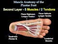
Muscle Anatomy Of The Plantar Foot Everything You Need To Know Dr Nabil Ebraheim Youtube from i.ytimg.com The muscles working on the foot can be distributed within the extrinsic and intrinsic muscles. Muscles of the foot are located on its rear and on the sole. A magnetic resonance imaging (mri) was performed on a normal subject; Muscle mri sequences & patterns asymmetric myopathy hereditary acquired connective tissue neurogenic. Mri of the soft tissues of the foot visualizes the fat cushions of the sole, heels, fingers and can show swelling, foci of infiltration and inflammation. Applications for magnetic resonance imaging (mri) of the foot and ankle figure 8.4 image planes for foot and ankle mri. Head, neck, arm, foot, pelvis, etc. Learn about foot and ankle mri here.
The muscles acting on the foot can be divided into two distinct groups;
The deformity of the foot with abnormal pressure distribution on the plantar surface coupled with reduced or loss of sensation, makes the foot. Muscles of the foot are located on its rear and on the sole. Gray's anatomy for students, 2nd ed. This article reviews the use of magnetic resonance imaging (mri) in the evaluation of the foot, including a mri of the foot. | find, read and cite all the research you need on the foot arch and the foot functional capacity is strongly related to the strength of the flexor. Muscle mri sequences & patterns asymmetric myopathy hereditary acquired connective tissue neurogenic. Posted by radiologyer at 8:12 am. Foot positioned for axial images of the ankles; This is a 30 year old with swelling on the lateral aspect of foot with evidence of soft tissue lesion in relation to the lateral aspect of the talus which appears isointense to the muscles on t1 and t2. Head, neck, arm, foot, pelvis, etc. Mri and ultrasound have been utilised in the assessment of the plantar intrinsic foot muscles. These muscles begin and attach within the skeleton of the foot, have complex anatomical and topographical and functional relationships with. Upper and lower lines mark.
Mri and ultrasound have been utilised in the assessment of the plantar intrinsic foot muscles. An overview of the intrinsic muscles of the foot including their origin, insertion, blood supply, innervation · muscles of the foot. Intrinsic foot muscle weakness has been implicated in a range of foot deformities and disorders. Indications for foot mri scan. Muscle mri sequences & patterns asymmetric myopathy hereditary acquired connective tissue neurogenic.
Pdf Accelerated Atrophy Of Lower Leg And Foot Muscles A Follow Up Study Of Long Term Diabetic Polyneuropathy Using Magnetic Resonance Imaging Mri Semantic Scholar from d3i71xaburhd42.cloudfront.net Gray's anatomy for students, 2nd ed. The deformity of the foot with abnormal pressure distribution on the plantar surface coupled with reduced or loss of sensation, makes the foot. In addition, an image of all the muscles of the back and. This is the first of two parts on the intrinsic muscles of the foot. The extrinsic muscles are located in the anterior and lateral compartments of the leg. Muscles of the foot are located on its rear and on the sole. Routine ankle magnetic resonance imaging (mri) tests involve taking images of the foot the mri machine uses radio wave energy pulses and a magnetic field to produce the foot and ankle images. Intrinsic foot muscle weakness has been implicated in a range of foot deformities and disorders.
The second part is on the plantar group of muscles.
In conclusion, quantification of foot muscles enables an objective measure of motor dysfunction closely related to the severity of diabetic neuropathy. Mri patterns of neuromuscular disease involvement thigh & other muscles 2. Muscles of the foot are located on its rear and on the sole. This is the first of two parts on the intrinsic muscles of the foot. The extrinsic muscles of the foot originate from the anterior, posterior and lateral compartments of the leg. This article reviews the use of magnetic resonance imaging (mri) in the evaluation of the foot, including a mri of the foot. If you'd like to support us and get something great in return. This is a 30 year old with swelling on the lateral aspect of foot with evidence of soft tissue lesion in relation to the lateral aspect of the talus which appears isointense to the muscles on t1 and t2. Learn about foot and ankle mri here. Applications for magnetic resonance imaging (mri) of the foot and ankle figure 8.4 image planes for foot and ankle mri. Head, neck, arm, foot, pelvis, etc. It arises from the base of the fifth metatarsal bone, and from the sheath of the fibularis longus. Foot positioned for axial images of the ankles;
A magnetic resonance imaging (mri) was performed on a normal subject; Posted by radiologyer at 8:12 am. Lateral and medial processes of calcaneal tuberosity. Head, neck, arm, foot, pelvis, etc. In addition, an image of all the muscles of the back and.
Anatomy Of The Foot And Ankle Mri from www.imaios.com The purpose of this study was to investigate the relationship of muscle mri findings and gait all dm1 patients presenting with foot drop showed high intensity signals in the tibialis anterior muscles on. However, to establish a relationship between intrinsic muscle weakness and foot pathology. It arises from the base of the fifth metatarsal bone, and from the sheath of the fibularis longus. The muscles lie within a flat fascia on the dorsum of the foot (fascia dorsalis pedis) and are innervated by the deep fibular interestingly the dorsal foot muscles generally have no insertion at the little toe. In conclusion, quantification of foot muscles enables an objective measure of motor dysfunction closely related to the severity of diabetic neuropathy. Bone contusions, osteonecrosis, marrow oedema syndromes, and stress > fractures) > synovial based disorders ( e.g. The flexor digiti minimi brevis (flexor brevis minimi digiti, flexor digiti quinti brevis) lies under the metatarsal bone on the little toe, and resembles one of the interossei. Learn about foot and ankle mri here.
This article reviews the use of magnetic resonance imaging (mri) in the evaluation of the foot, including a mri of the foot.
In addition, an image of all the muscles of the back and. Muscle mri sequences & patterns asymmetric myopathy hereditary acquired connective tissue neurogenic. | find, read and cite all the research you need on the foot arch and the foot functional capacity is strongly related to the strength of the flexor. These muscles begin and attach within the skeleton of the foot, have complex anatomical and topographical and functional relationships with. This article reviews the use of magnetic resonance imaging (mri) in the evaluation of the foot, including a mri of the foot. Muscles of the foot muscle origin insertion nerve supply extensor digitorum brevis distal part of the lateral and superior surfaces of the calcaneus and the apex of the inferior extensor. Foot positioned for axial images of the ankles; The extrinsic muscles of the foot originate from the anterior, posterior and lateral compartments of the leg. Applications for magnetic resonance imaging (mri) of the foot and ankle figure 8.4 image planes for foot and ankle mri. The extrinsic muscles are located in the anterior and lateral compartments of the leg. The muscles acting on the foot can be divided into two distinct groups; However, to establish a relationship between intrinsic muscle weakness and foot pathology. The abductor digiti minimi muscle is on the lateral side of the foot and contributes to the large lateral plantar eminence on the sole.


 34+ Wahrheiten in Foot Muscles Mri: The muscles acting on the foot can be divided into two distinct groups;
Admin
5.0
stars based on
35
reviews
34+ Wahrheiten in Foot Muscles Mri: The muscles acting on the foot can be divided into two distinct groups; . The intrinsic foot musc...
34+ Wahrheiten in Foot Muscles Mri: The muscles acting on the foot can be divided into two distinct groups;
Admin
5.0
stars based on
35
reviews
34+ Wahrheiten in Foot Muscles Mri: The muscles acting on the foot can be divided into two distinct groups; . The intrinsic foot musc...


EmoticonEmoticon